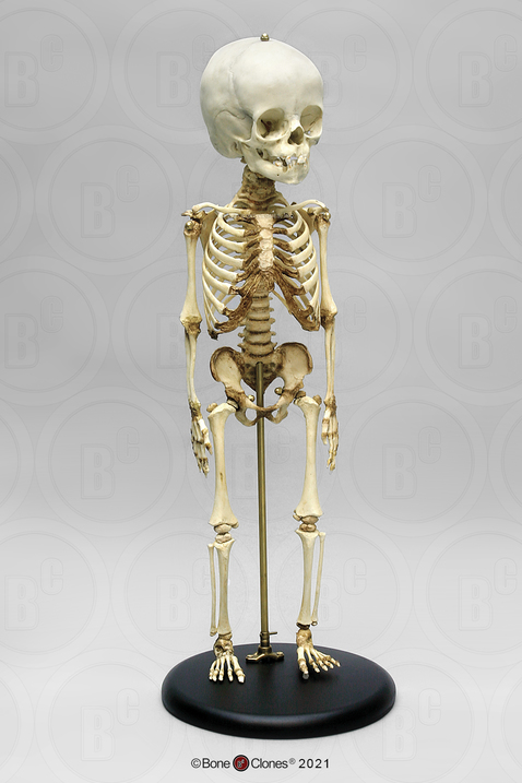
Child girl skeleton anatomy Royalty Free Vector Image Biology Diagrams This seeming paradox is rooted in the fact that there is intense skeletal growth and development during childhood and adolescence, and much more bone is formed than lost. Later in life, the loss of bone tissue exceeds the rate of bone replacement. It, therefore, follows that lifelong bone health is dependent on maximizing peak bone mass during

They occur at 6-month intervals of skeletal age from 11 years in girls and 13 in boys. The fully fused olecranon occurs at skeletal age of 13 in girls and 15 in boys and marks the deceleration of growth velocity. 9. Determination of skeletal age using the Modified Tanner-Whitehouse III Skeletal Maturity Assessment (Table 1.2 and Fig. 1.12) Purpose of review: The purpose of this review is to summarize the recent clinical findings surrounding the muscle-bone relationships in children, while considering muscle adiposity, endocrine factors, and lifestyle influences (i.e., diet and exercise) involved in pediatric musculoskeletal development. Recent findings: Positive relationships between cortical bone geometry and muscle mass, size A child's chronological age, determined by their birthdate, does not always align with their skeletal age, which reflects the biological maturity of their bones. While most children follow a predictable growth pattern, variations in skeletal development can reveal important insights about overall health and future growth potential.
Musculoskeletal health in children and adolescents Biology Diagrams
Children have growth plates in each long bone. A growth plate is an area of soft bone at each end of the long bones. Growth plates allow the bone to grow as the child grows. The growth plates fuse by the time a child is 14 to 18 years old. Skeletal muscle and pediatric bone development. Curr Opin Endocrinol Diabetes Obes (2015) 22:467-74. doi: 10.1097/MED.0000000000000201 [Google Scholar] 3. Fornari ED, Suszter M, Roocroft J, Bastrom T, Edmonds EW, Schlechter J. Childhood obesity as a risk factor for lateral condyle fractures over supracondylar humerus fractures.

An adequate development of skeletal muscles during these two periods may have long-term consequences on body composition and inclination to engage in physical activity throughout life . Moreover, although muscle fiber composition is genetically determined ( 8 ), early physical training can play an important role in "fiber reprogramming" ( 9 ).

Skeletal muscle and pediatric bone development Biology Diagrams
The musculoskeletal system is influenced by many different factors as infants and children grow. It can adapt to the demands, or lack of demands, that are placed on it. The major load on bone comes from muscle forces. When muscle pull is altered due to genetic or neuromuscular conditions, alignment may be impacted. Atypical alignment can directly affect functional activities and an individual
